
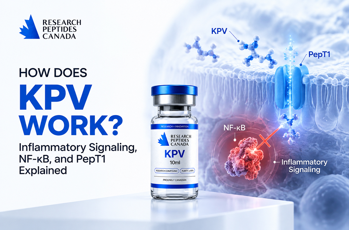


Understanding how does KPV work requires looking beyond its small size.
KPV peptide is a three-amino-acid peptide derived from the C-terminal sequence of alpha-melanocyte-stimulating hormone (α-MSH). Despite containing only lysine, proline, and valine, research suggests that KPV can influence several inflammatory pathways involved in immune signaling, epithelial responses, and cellular stress.
The most studied mechanisms involve:
- regulation of NF-κB inflammatory signaling;
- transport through the peptide transporter PepT1;
- reduction of pro-inflammatory cytokine activity;
- modulation of oxidative stress responses.
However, most evidence comes from cell and animal models. While these studies provide important mechanistic insight, they do not yet establish how KPV behaves as a therapeutic intervention in humans.
If you are exploring peptide compounds for laboratory research, visit Research Peptides Canada for research-use-only compounds and available documentation.
How Does KPV Work at the Cellular Level?
The foundation of how does KPV work begins with its origin from α-MSH.
Alpha-melanocyte-stimulating hormone is part of the melanocortin peptide family and is produced from the precursor protein pro-opiomelanocortin (POMC).
Although full-length α-MSH is known for regulating pigmentation and immune responses, researchers discovered that its final three amino acids—lysine, proline, and valine—retain important anti-inflammatory activity.
Early research demonstrated that KPV could reduce inflammatory responses even when classical melanocortin receptor signaling was disrupted.
In a mouse inflammation model, Getting and colleagues found that KPV reduced inflammatory leukocyte accumulation and maintained activity even in animals lacking functional melanocortin-1 receptors (MC1R). This suggested that KPV may operate through mechanisms different from traditional α-MSH signaling (Getting et al., 2003, Journal of Endocrinology).
This discovery changed how researchers viewed KPV.
Rather than acting simply as a smaller version of α-MSH, KPV appeared to function as an independent signaling molecule capable of influencing inflammatory pathways directly.
At the cellular level, current research suggests that KPV may:
- enter specific cell types through peptide transport systems;
- interfere with inflammatory transcription pathways;
- reduce production of inflammatory mediators;
- influence oxidative stress-related signaling.
The exact receptor or molecular target responsible for all KPV effects remains an active area of investigation.
For researchers interested in exploring KPV further, learn more about KPV 10mg and its research applications.
How Does KPV Influence NF-κB and Inflammatory Signaling?
One of the most important explanations for how does KPV work involves the NF-κB pathway.
NF-κB is a transcription factor that controls the expression of many inflammatory genes.
When activated by signals such as:
- bacterial products;
- inflammatory cytokines;
- oxidative stress;
- tissue injury;
NF-κB moves into the nucleus and stimulates production of inflammatory mediators.
These include:
- TNF-α;
- IL-1β;
- IL-6;
- IL-8;
- other immune-response molecules.
Research suggests that KPV can suppress this inflammatory cascade.
A key study by Dalmasso and colleagues investigated KPV in intestinal epithelial and immune cells. Researchers found that KPV inhibited NF-κB activation, reduced inflammatory cytokine production, and decreased MAP kinase signaling pathways involved in intestinal inflammation (Dalmasso et al., 2008, Gastroenterology).
The study also showed that KPV reduced activation of inflammatory pathways without relying on classical melanocortin receptor activation.
This is important because it suggests that KPV may regulate inflammation through a different biological route compared with α-MSH.
Further research in airway epithelial cells supported this mechanism.
Moustafa and colleagues found that KPV reduced TNF-α-induced NF-κB activity, decreased IL-8 production, and reduced inflammatory signaling associated with airway epithelial responses (Moustafa et al., 2012).
Together, these findings provide one of the strongest explanations for how KPV works:
KPV enters inflammatory cells → reduces NF-κB activation → decreases inflammatory mediator production.
However, the exact molecular interaction between KPV and NF-κB signaling is still being investigated.
What Role Does PepT1 Play in KPV Activity?
One of the most interesting discoveries in KPV research is the role of PepT1, or peptide transporter 1.
PepT1 is a transporter normally responsible for absorbing small dietary peptides and amino acids in the intestine.
Under normal conditions, PepT1 expression in intestinal epithelial cells is relatively controlled.
During inflammation, however, PepT1 expression can increase.
This creates a potential pathway for small peptides such as KPV to enter intestinal cells.
Dalmasso and colleagues demonstrated that intestinal epithelial cells transported KPV through PepT1. Blocking PepT1 reduced KPV uptake and weakened its anti-inflammatory effects, suggesting that PepT1 is an important part of KPV activity in intestinal models (Dalmasso et al., 2008, Gastroenterology).
This discovery helped explain why KPV has received particular attention in gut inflammation research.
The proposed mechanism is:
- Intestinal inflammation increases PepT1 expression.
- PepT1 transports KPV into epithelial or immune cells.
- KPV influences inflammatory signaling pathways.
- NF-κB activation and cytokine production decrease.
However, this mechanism is highly dependent on tissue environment.
PepT1 expression varies between tissues, meaning the same transport mechanism may not occur equally throughout the body.
How May KPV Affect Cytokines and Oxidative Stress?
Beyond NF-κB, researchers have investigated how KPV influences cytokines and oxidative stress responses.
Inflammation involves complex communication between immune cells and tissues.
Cytokines act as signaling molecules that coordinate immune activity. Excessive cytokine production can contribute to chronic inflammatory states.
Experimental studies suggest that KPV may reduce several inflammatory cytokines, including:
- IL-1β;
- TNF-α;
- IL-8.
In intestinal inflammation models, KPV reduced inflammatory markers and improved tissue-related outcomes.
Kannengiesser and colleagues tested KPV in mouse models of inflammatory bowel disease and found reductions in inflammatory infiltration, myeloperoxidase activity, and disease severity. These effects occurred even in models with impaired MC1R signaling, supporting an MC1R-independent mechanism (Kannengiesser et al., 2008, Inflammatory Bowel Diseases).
More recent research has expanded into oxidative stress.
A 2025 study examined KPV in human HaCaT keratinocytes exposed to particulate matter-induced stress. Researchers found that KPV reduced reactive oxygen species generation, decreased inflammatory signaling through MAPK and NF-κB pathways, and reduced markers associated with inflammatory cell death. The study also confirmed protective effects in a three-dimensional skin model (KPV keratinocyte study, 2025).
These findings suggest that KPV may influence inflammation not only by reducing cytokines but also by modifying cellular stress responses.
However, these effects remain experimental.
For researchers interested in exploring KPV further, learn more about KPV 10mg and its research applications.
Does the KPV Mechanism Observed in Preclinical Research Translate to Humans?
This is the most important unanswered question in KPV peptide research.
The current mechanism is supported by:
- intestinal cell studies;
- immune-cell experiments;
- animal inflammation models;
- skin-cell models.
However, direct human intervention data remain extremely limited.
The U.S. FDA has noted that it has not identified clinical studies or human exposure data for KPV through any route of administration. The agency also highlighted uncertainty regarding safety, pharmacology, and human biological effects (U.S. FDA, 2026, KPV scientific review).
This means researchers still do not know:
- how KPV is absorbed in humans;
- how long it remains biologically active;
- which tissues receive meaningful exposure;
- whether animal anti-inflammatory effects occur in humans;
- what long-term safety profile exists.
Another challenge is delivery.
Because KPV is a small peptide, researchers are studying different delivery approaches.
A transdermal delivery study found that passive skin penetration of KPV was limited, while technologies such as microneedles and iontophoresis improved peptide transport through skin models (Babu et al., 2017).
This highlights an important principle:
A biological effect observed in a laboratory system depends not only on the peptide itself, but also on whether enough active compound reaches the target tissue.
What About Research Dosing?
There is currently no validated human dosing protocol for KPV.
Published concentrations are experimental parameters.
For example:
- intestinal studies used laboratory concentrations ranging from nanomolar levels;
- skin-cell research used concentrations such as 50 μg/mL;
- animal studies used model-specific exposures.
These values cannot be directly converted into human dosing because absorption, metabolism, distribution, and biological exposure differ significantly between experimental systems.
Learn more in our complete guide: KPV Peptide: What It Is and What Current Research Actually Shows.
FAQ About How KPV Works
Is KPV derived from α-MSH?
Yes. The KPV peptide represents the final three amino acids of α-MSH: lysine, proline, and valine.
Does KPV activate melanocortin receptors?
Not necessarily. Research suggests KPV can maintain anti-inflammatory activity even when melanocortin receptor signaling is impaired, indicating additional mechanisms (Getting et al., 2003).
What is the main mechanism of KPV?
The strongest evidence suggests that KPV influences inflammatory signaling by reducing NF-κB activation and regulating cytokine production.
Why is PepT1 important for KPV?
PepT1 may transport KPV into intestinal cells, especially during inflammatory conditions when PepT1 expression increases.
Does KPV reduce inflammation in humans?
Currently, human evidence is insufficient. Most findings come from experimental models.
Is there an approved KPV dose?
No. There is currently no established clinical dosing protocol for KPV.
Overall, understanding how does KPV work requires looking at several connected mechanisms rather than one single pathway.
Current research suggests that KPV acts as an α-MSH-derived signaling peptide capable of influencing NF-κB activity, PepT1-dependent transport, cytokine regulation, and oxidative stress responses.
The strongest evidence remains preclinical, particularly in intestinal inflammation and epithelial models.
Future human studies will be needed to determine whether these mechanisms translate into meaningful biological effects in people.
For laboratory-focused peptide research, visit Research Peptides Canada to explore research-use-only compounds and available documentation.
Disclaimer: The information and products discussed on this website are intended strictly for laboratory research and educational purposes only. They are not intended for human or veterinary use, diagnosis, treatment, prevention, or any form of clinical application.
3 Comments
I really liked how clearly this article explains the connection between PepT1 transport and KPV’s activity in intestinal models. The fact that transport into cells may influence the observed anti-inflammatory effects adds an interesting layer to the mechanism. I’d be curious to see whether researchers identify similar transport pathways in other tissues.
The section on NF-κB was particularly helpful because it connects the cellular mechanism with changes in inflammatory signaling. I also appreciate that the article doesn’t present the preclinical findings as proof of human effects. It will be interesting to see whether future studies can clarify how these pathways behave in human models.
This was a great overview of how KPV may influence more than one inflammatory pathway at the same time. The discussion around cytokines, oxidative stress, and tissue-specific delivery makes the current research picture much more nuanced. I’d be interested to see which of these mechanisms receives the most attention in future human research.